38 labeled cow eye dissection
Cow Eye Labeling Quiz - PurposeGames.com About this Quiz. This is an online quiz called Cow Eye Labeling. There is a printable worksheet available for download here so you can take the quiz with pen and paper. PDF Cow Eye Dissection - University of Illinois Inform the students that there are cow eye dissection guides on the dissection tables and they should follow the directions in the guide so they can appropriately identify structures on the exterior and interior of the eye. Activity 1: Examination of the exterior of the eye Start by having the students examine the exterior of the eye.
Cow Eye Dissection Worksheet Answer - Ehydepark Below is an example of a response for. (optic nerve, iris, pupil, sclera, cones, rods, cornea, retina, lens and vitreous humor) use a labeled drawing if it is helpful. Answer the worksheet questions on the cow eye dissection. Locate the covering over the front of the eye, the cornea. Materials • preserved cow eye • scalpel or scissors ...

Labeled cow eye dissection
Cow Eye Dissection Quiz - PurposeGames.com cow eye, dissection Remaining 0. Correct 0. Wrong 0. Press play! 0%. 0:00.0. Quit. Again. This game is part of a tournament. You need to be a group member to play the tournament. Join group, and play Just play. Your Scorecard. The scorecard of a champion. Score . 0 % Time . 0:00.0. Place # 0. Lab: Cow Eye Dissection Flashcards | Quizlet Lab: Cow Eye Dissection STUDY Flashcards Learn Write Spell Test PLAY Match Gravity Created by Daniel_Kinsey Terms in this set (14) Anterior Chamber Ciliary Muscle enabling changes in lens shape for light focusing Retina convert the light into neural signals, and send these signals on to the brain for visual recognition Choroid PDF Cow Eye Dissection - mbusd.org • Preserved cow's eye • Forceps • Dissection probes • Dissection scissors • Dissection tray • Vinyl or latex gloves • Apron or tee-shirt • Paper towels • Plastic trash bag External features of the eye A. Locate the cornea, sclera, and optic nerve. a. The white part of the eye, the sclera, is a tough, outer covering
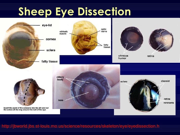
Labeled cow eye dissection. PDF COW'S EYE dissection - Exploratorium COW'S EYE dissection page 6 Now take a look at the rest of the eye. If the vitreous humor is still in the eyeball, empty it out. On the inside of the back half of the eyeball, you can see some blood vessels that are part of a thin fleshy film. That film is the retina. Before you cut the eye open, the vitreous humor PDF Cow Eye Dissection: Examining Structure and Function During this activity, you will dissect a cow eye. You will observe several important features of the eye and develop your understanding of how each part functions to make vision possible. Materials • Preserved Cow Eye • Scalpel or Scissors • Forceps • Dissection Tray • Gloves • Safety Glasses • Lab Apron 1. Cow Eye Dissection - YouTube About Press Copyright Contact us Creators Advertise Developers Terms Privacy Policy & Safety How YouTube works Test new features Press Copyright Contact us Creators ... Eye Dissection Lab.pdf - Student: Jacqueline Labeled... Labeled cow eye dissection; Far Eastern University • BIO 1225. LAB ACTIVITY_Eye dissection.pdf. 1. BIO 160 5 week SUMMER Unit 3 Schedule.docx. Horizon High School, Scottsdale. BIO 160. cardiovascular system; Blood Pressure Lab; BIO160 Schedule; Horizon High School, Scottsdale • BIO 160.
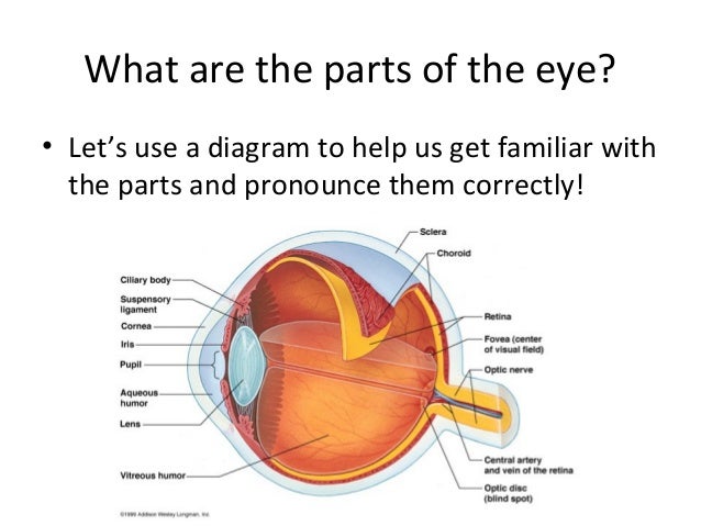
Cow Eye Dissection Teaching Resources | Teachers Pay Teachers Cow Eye Dissection by CrazyScienceLady 49 $5.00 PDF Cow Eye Dissection: Directions and questions for dissecting a cow eye. Also includes a diagram of the eye, fun facts, an activity on how to find your blind spot and all answer keys. Appropriate for grades 4-7, I run this lab for each of the 4th grade classes at my school. It's a HUGE hit!!! Cow Eye Dissection - PBS LearningMedia This collection details the anatomy of a cow eye. Cow Eye Dissection Parts Labeled - All About Cow Photos Solved 6 The Images Below Show A Preserved Cow S Eye One Chegg Cow Eye Dissection Google Slides Dissection Cattle Anatomy Human Eye Png Clipart Biology Blue Glow Diagram Neur 320 Art And Vision 3 Use The Pictures Below To Name Parts Of Eye That You Will Observe In Course Hero Eye Dissection Cow S Eye Dissection Diagram Cow Eye Dissection & Parts of the Eye Diagram | Quizlet cornea Clear, outer layer of the front of the eye. sclera White, outermost layer of the eye. Helps maintain shape and gives attachment to muscles. photoreceptors The cells in the retina that respond to light (rods and cones) rods Photoreceptor cells in the eye that detect black, white, and gray cones Photoreceptor cells in the eye that detect color
PDF Cow Eye Dissection Lab This cow eye dissection kit comes with everything you need to conduct a lab examination. Safety Guidelines • Work in a place separate from eating and food preparation areas. • Use disposable latex gloves or nitrile gloves during the dissection and cleanup. • Use only dissection tools provided. Cow's Eye Dissection - Eye diagram - Exploratorium A muscle that controls how much light enters the eye. It is suspended between the A cow's iris is brown. many colors, including brown, blue, green, and gray. A clear fluid that helps the cornea keep its rounded shape. The pupil is the dark circle in the center of your iris. It's a hole that Your pupil is round. Cow Eye Dissection | Carolina.com Students explore the external and internal anatomy, learning how structures work together to create images from incoming light. A preserved cow eye dissection can be carried out in 1-2 class periods and only requires basic dissecting instruments. Explore the internal and external anatomy of the cow eye using the procedural steps below. Cow Eye Dissection - The Biology Corner COW EYE DISSECTION 1. Examine the outside of the eye. You should be able to find the sclera, or the whites of the eye. This tough, outer covering of the eyeball has fat and muscle attached to it 2. Locate the covering over the front of the eye, the cornea. When the cow was alive, the cornea was clear.
Dissecting An Eyeball - Krieger Science Returning to our dissection, underneath the retina is a pretty, shiny, blue-green mirror, officially called the tapetum. This is what causes cow's eyes to shine in headlights, and it is probably the most memorable part of an eye dissection for children. This is also one thing that cows and people do not have in common.
PDF Cow Eye Dissection Guide - Central Bucks School District DISSECTION OF THE COW EYE Please make sure to wear gloves and safety glasses when you are dissecting, and make sure to clean up thoroughly after the lab. Also, the cow eyes can be rather slippery, so use caution when handling and cutting them. You will need a scalpel and forceps. 1. First, identify the most external structures of the eye.
Cow Eye Dissection Lab from Anatomy and Physiology Cow's Eye Dissection Lab Page 2. List the two functions of the lens. The lens is a transparent structure in the eye that, along with the cornea, helps to refract and focus light. To help us focus on closer objects is gets fatter to refract more light and vice versa for focusing on. objects further away.
Cow Eye Dissection Guide Each fragile part of the eye works together to provide information to the brain, and the brain interprets it instantaneously giving you a perfect image. It is an amazing process. Download: Cow Eye Dissection Lab. A cow's eye, like other farm animal organs, is similar to our eyes. One benefit of a cow eye dissection is that by examining the
PDF Name: Dissection 101: Cow Eye human eye _____ _____ Draw and label the cow eye. Cornea Optic nerve Vitreous humor Retina Optic disc (blind spot) Choroid Tapetum lucidum Sclera Aqueous humor Suspensory ligaments Lens Ciliary body Pupil Iris Provided by Dissection 101: Cow Eye
Cow Eye Dissection Guide - Google Slides Cow Eye. Use the point of a scissors or a scalpel to make an incision through the layers of the eye capsule (similar to figure 1); there are three layers from the exterior: sclera, whitish/grey, continuous with the transparent cornea, choroid, thin dark black layer and the retina, thin greyish/pink layer. Use a scissors to dissect the entire ...
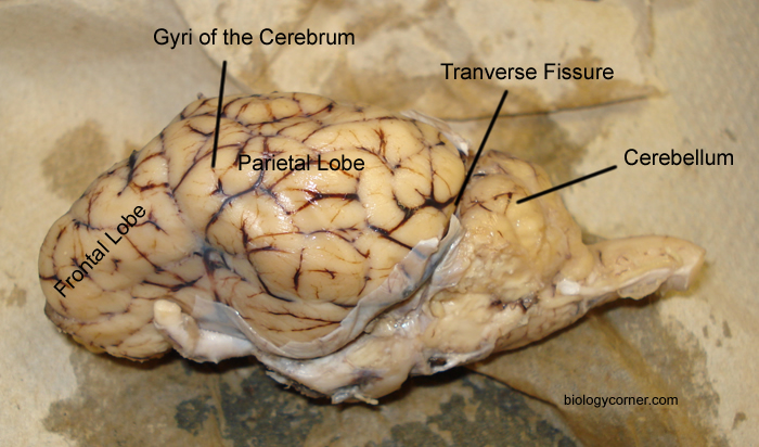
Cow Eye and Sheep Brain Dissection Report - Prezi By dissecting and examining the anatomy of a preserved cow eye, you can learn how your own eye forms images and sends these images to your brain. Introduction The sheep brain and the cow eye are used in the study of human anatomy because they are structurally similar to that of a human specimen.
Cow eye - dissection and label Cow eye shown with labeled cornea. The cornea is the transparent front part of the eye that covers the iris, pupil, and anterior chamber. The cornea, with the anterior chamber and lens, refracts light, with the cornea accounting for approximately two-thirds of the eye's total optical power. 3.
Cow Eye Dissection Pre-lab Key - Google Docs 1. Use the structures listed in question #2 and label the diagram. 2. Describe the function of the following structures: Cornea - tough covering over the iris that helps protect the eye and directs the light towards the lens. Optic nerve- The bundle of nerve fibers that carries the impulse to the brain.


Post a Comment for "38 labeled cow eye dissection"