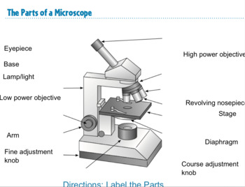
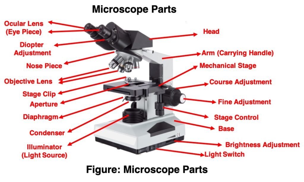
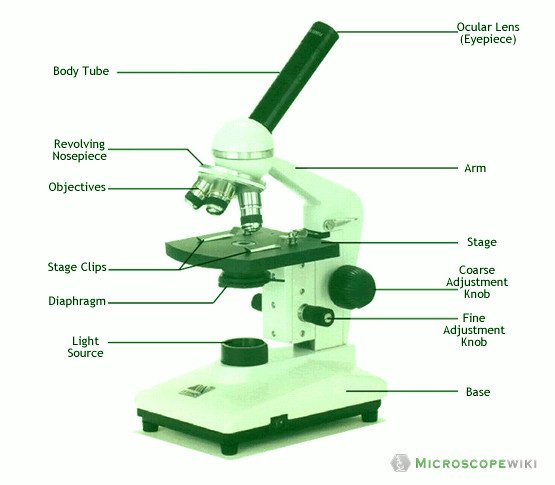
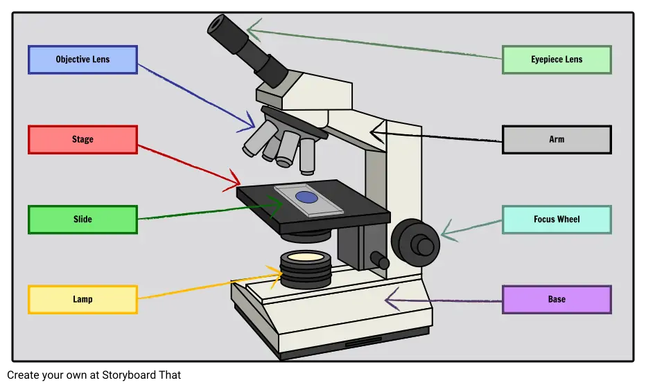
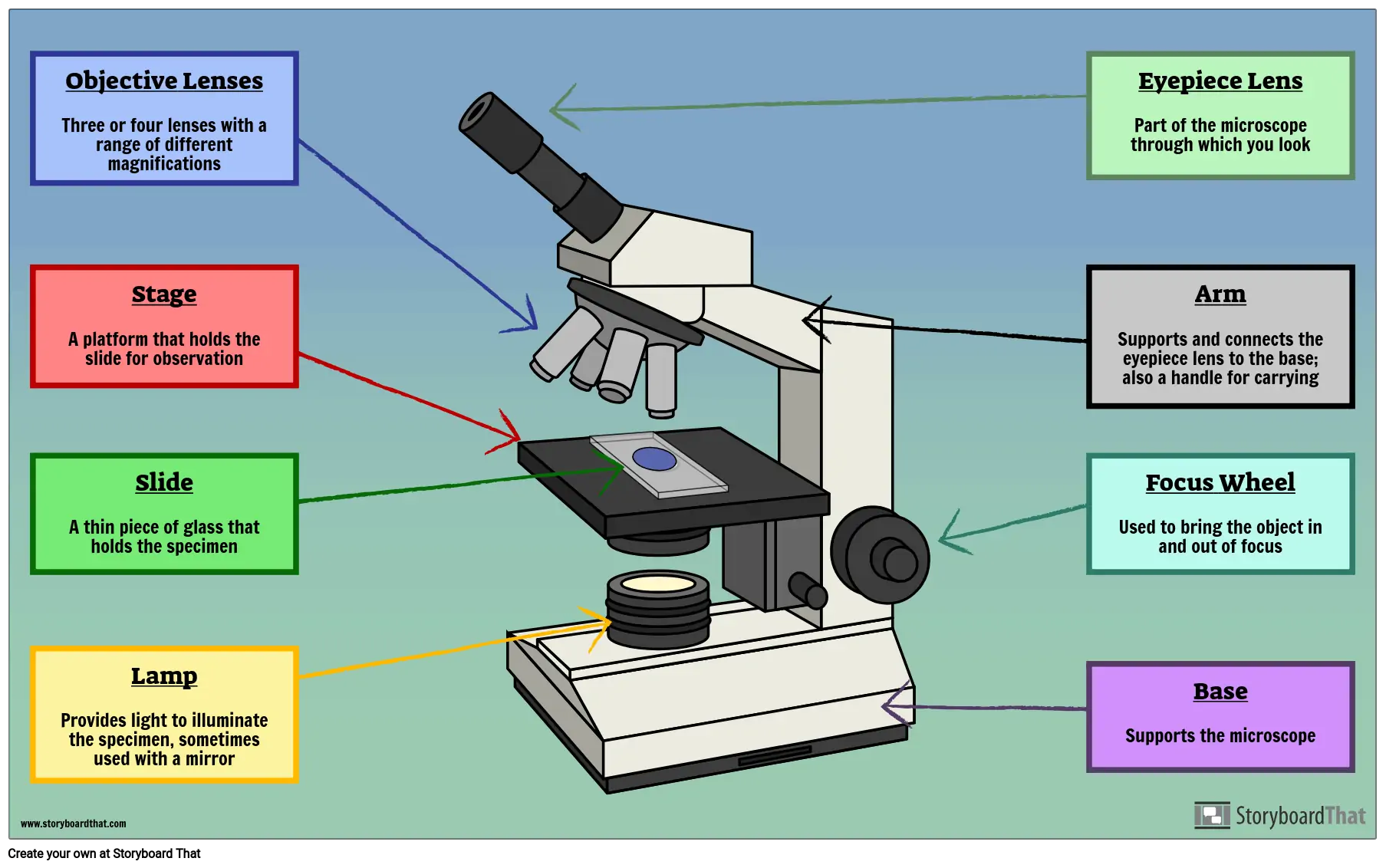
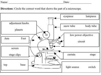
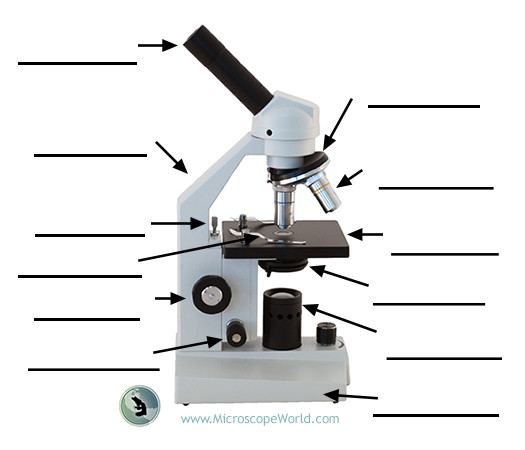
42 microscope labeled diagram
Variation in the fruit development gene POINTED TIP regulates ... The haploblocks are represented by inverted triangles. The darkness of the color of each box indicates an r2 value indicated in the color scale. The red star indicates the lead SNP containing... BIOLOGY FORM ONE NOTES FREE - Educationnewshub.co.ke Lenses are found in microscope and the hand lens (magnifier). Its frame is marked e.g. x8 or x10—indicating how much larger will be the image compared to object. Precautions during Collection and Observation of specimens Collect only the number of specimen you need. Do not harm the specimens during the capture or collection exercise.
Gram Staining Procedure | New Health Advisor 2. Label the Slides Draw a circle under the slides using a marking pen designed for glassware. This will help to designate which area to prepare the smear in the following step. You can also label them with the organism's initials at the edge of each slide. Take care that the labels do not get in contact with the reagentsused forstaining. 3.

Microscope labeled diagram
Light Microscope (Theory) - Amrita Vishwa Vidyapeetham Microscope is an optical instrument that uses lens or combination of lens to produce magnified images that are too small to seen by unaided eye. Microscope provides the enlarged view that helps in examining and analyzing the image. Compound Microscope- Definition, Labeled Diagram, Principle, … 03/04/2022 · Magnification of compound microscope. In order to ascertain the total magnification when viewing an image with a compound light microscope, take the power of the objective lens which is at 4x, 10x or 40x and multiply it by the power of the eyepiece which is typically 10x. BIO 240 SYLLABUS FALL 2022 2.doc - NATURAL SCIENCES... Digestive System: Anatomy 24 621 - 648 Models, Microscope slides & Dissection of Fetal pig 4. Digestive System: Physiology of Enzyme Action 24 649 - 650 Carbohydrate digestion 650 - 651 Protein digestion 652 - 653 Lipid digestion 653 - 662 5. Urinary System: Anatomy 25 663 - 688 Models, Microscope slides & Sheep kidney dissection 6.
Microscope labeled diagram. ECLIPSE Ti2 Series | Inverted Microscopes | Nikon Microscope Products ... Application Notes Leading platform for advanced imaging. The ECLIPSE Ti2 inverted microscope delivers an unparalleled 25mm field of view (FOV) that revolutionizes the way you see. With this incredible FOV, the Ti2 maximizes the sensor area of large-format CMOS cameras without making compromises, and significantly improves data throughput. Skin: Cells, layers and histological features | Kenhub It is comprised of three major layers: epidermis, dermis and hypodermis, which contain certain sublayers. Owing to variations in height and weight, the surface area of the skin may vary based on these parameters. The surface of the skin is a parameter that is often used in determining the therapeutic dose for various medications. Contents The Equine Lymphatic System and Treatment of Equine Chronic Progressive ... Risse, M. Concerning Pathogensis of Acute Lymphangitis in the Horse - A Histological, Immunohistological and Transition Electron Microscope Study. Inaugural Dissertation, The Veterinary University of Hannover, Vet, med. thesis, (2004) Rotting, A. et al Manual Lymph Drainage in the Horse for the Treatment of the Hind Limb. en.wikipedia.org › wiki › Electron_microscopeElectron microscope - Wikipedia An electron microscope is a microscope that uses a beam of accelerated electrons as a source of illumination. As the wavelength of an electron can be up to 100,000 times shorter than that of visible light photons , electron microscopes have a higher resolving power than light microscopes and can reveal the structure of smaller objects.
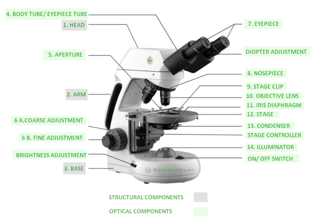
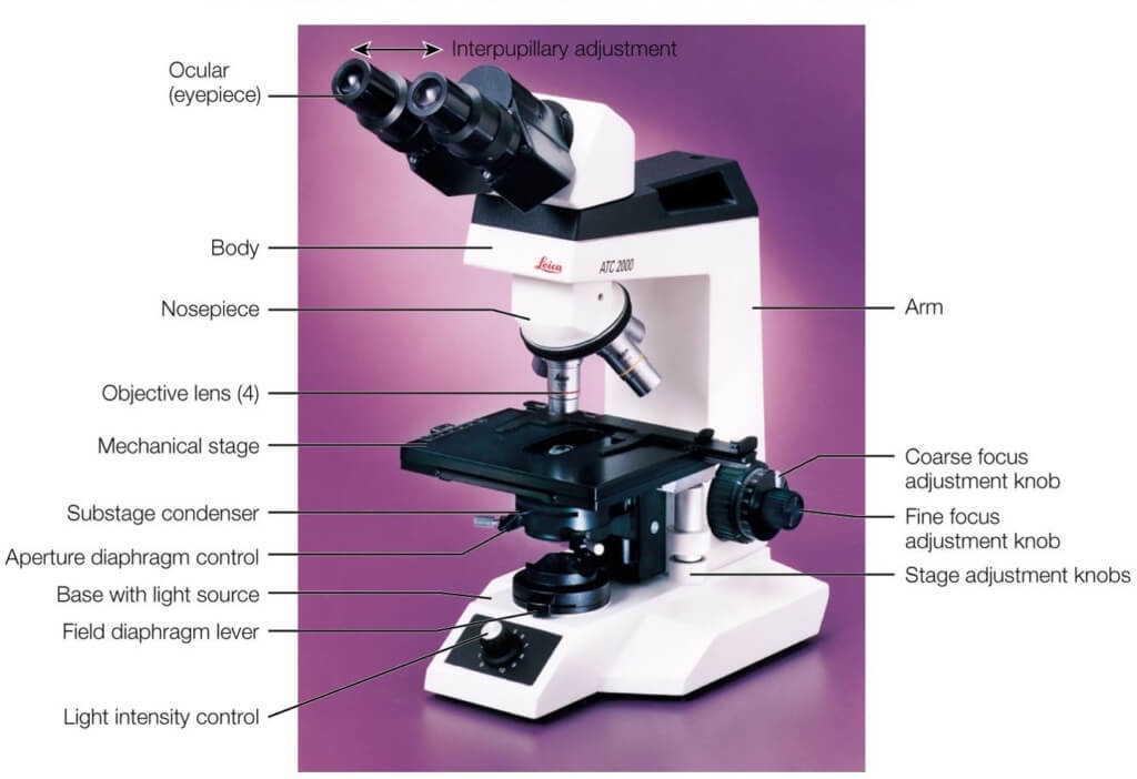
microbenotes.com › parts-of-a-microscopeParts of a microscope with functions and labeled diagram Sep 17, 2022 · Light Microscope- Definition, Principle, Types, Parts, Labeled Diagram, Magnification Amazing 27 Things Under The Microscope With Diagrams Plant Cell- Definition, Structure, Parts, Functions, Labeled Diagram Drosophila melanogaster - Wikipedia Drosophila melanogaster is a species of fly (the taxonomic order Diptera) in the family Drosophilidae.The species is often referred to as the fruit fly or lesser fruit fly, or less commonly the "vinegar fly" or "pomace fly". Starting with Charles W. Woodworth's 1901 proposal of the use of this species as a model organism, D. melanogaster continues to be widely used for biological research in ... microscopeinternational.com › compound-microscopeCompound Microscope Parts, Functions, and Labeled Diagram Nov 18, 2020 · Base: Bottom base of the microscope that houses the illumination & supports the compound microscope. Objective lenses: There are usually 3-5 optical lens objectives on a compound microscope each with different magnification levels. 4x, 10x, 40x, and 100x are the most common magnifying powers used for the objectives. Gram Stain Technique (Theory) : Microbiology Virtual Lab I ... It is also a key procedure in the identification of bacteria based on staining characteristics, enabling the bacteria to be examined using a light microscope. The bacteria present in an unstained smear are invisible when viewed using a light microscope. Once stained, the morphology and arrangement of the bacteria may be observed as well.
Multipolar Neurons - Structure, functions and diagram - GetBodySmart Multipolar neurons have three or more processes attached to the cell bodies. 1. 2. One process serves as the axon, which conducts electrochemical impulses ( action potentials) between cells. 1. 2. The remaining processes are dendrites. Together, the cell body and dendrites form the receptive zone of multipolar neurons. 1. What is LabVIEW? Graphical Programming for Test & Measurement - NI LabVIEW Base. Recommended for building simple test and measurement applications. Includes the standard capabilities of LabVIEW: Acquire data from NI and third-party hardware and communicate using industry protocols. Create interactive UIs for test monitoring and control. Utilize standard math, probability, and statistical functions. rsscience.com › compound-microscope-parts-labeledCompound Microscope Parts – Labeled Diagram and their ... There are three major structural parts of a microscope: Head, Base, and Arm. Always lift a microscope by holding both the arm and base with two hands. There are two major optical lens parts of a microscope: Eyepiece (10x) and Objective lenses (4x, 10x, 40x, 100x). Darkfield Microscope- Definition, Principle, Uses, Diagram 13/03/2022 · Parts of a microscope with functions and labeled diagram; 22 Types of Spectroscopy with Definition, Principle, Steps, Uses; Animal Cell- Definition, Structure, Parts, Functions, Labeled Diagram; Limitations of Darkfield Microscope. The main limitation of dark-field microscopy is the low light levels seen in the final image. The sample must be very …
microbenotes.com › compound-microscope-principleCompound Microscope- Definition, Labeled Diagram, Principle ... Apr 03, 2022 · Parts of a microscope with functions and labeled diagram Light Microscope- Definition, Principle, Types, Parts, Labeled Diagram, Magnification Amazing 27 Things Under The Microscope With Diagrams
onion | Description, History, Uses, Products, Types, & Facts onion, (Allium cepa), herbaceous biennial plant in the amaryllis family (Amaryllidaceae) grown for its edible bulb. The onion is likely native to southwestern Asia but is now grown throughout the world, chiefly in the temperate zones. Onions are low in nutrients but are valued for their flavour and are used widely in cooking. They add flavour to such dishes as stews, roasts, soups, and salads ...
What is the Largest Biological Cell? (with pictures) - All the Science The largest biological cell is often cited as the ostrich egg, which is about 6 inches (15 cm) long and weigh about 3 pounds (1.4 kg). This is a myth. There are at least several biological cells larger than an ostrich egg, despite the fact that even many scientists and laypeople believe the ostrich egg is indeed the biggest.
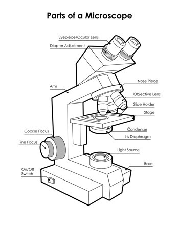
slidingmotion.com › microscope-parts-functionMicroscope Parts, Function, & Labeled Diagram - slidingmotion To examine these small objects with high magnification, parts of a microscope are made with special components with high accuracy. Due to that, accurate examination and results are possible to achieve. Microscope parts labeled diagram gives us all the information about its parts and their position in the microscope. Microscope Parts Labeled Diagram
Home | MyRSU - Rogers State University Benefit Open Enrollment is October 17-28, 2022! All benefit-eligible employees are required to participate. Log in to MyRSU to either enroll or decline benefits & upload proof of alternative medical coverage. No plans or providers are changing for 2023. Providers' 2023 plans & rate sheets will be posted on MyRSU soon for your review.
Mr. Jones's Science Class Matter: Atoms and Properties - Open Response Question 3. Force and Motion - Open Response Question 3. Forms of Energy - Open Response Question 1. Forms of Energy - Open Response Question 2. Earth's Structure & Natural Processes - Open Response Question 1.
Welcome to Boreal Science Printed from Boreal Science Website User: [Anonymous] Date: 10-10-2022 Time: 07:32
X-Ray Generation Notes - University of Oklahoma At low resolution (lower scattering angle) the K α wavelength is considered as a weighted average of the K α 1 and K α 2 lines with λ ( K α ave) = [2* (λ ( K α 1 )) + λ ( K α 2 )]/3. The K α line is about 5 - 10 times as intense as the K β line. The intensity of the K α line can be approximately calculated by I k = B i (V - V k) 1.5
Spleen histology: Location, functions, structure | Kenhub Spleen histology slide (labeled) The spleen is a fist sized organ located in the left upper quadrant of the abdomen. It is the largest lymphoid organ and thus the largest filter of blood in the human body. The spleen has a unique location, embryological development and histological structure that differs significantly from other lymphoid organs.
Animal Cell Model with Labeled Parts - homesciencetools.com Visualize a cell's parts with this sturdy soft foam model. One half has labeled parts: mitochondria, smooth and rough ER, lysosomes, Golgi apparatus, vacuoles, centrioles, ribosomes, cytoplasm, cell membrane, and nucleus with nucleolus and chromatin. The other half has the same parts, only unlabeled for testing.
Lighting Ergonomics - Survey and Solutions : OSH Answers The amount of light falling on a surface is measured in units called lux. Depending on the factors noted above, adequate general lighting is usually between 500 and 1000 lux when measured 76 cm (30 inches) above the floor.*. Examples of industrial and office tasks and the recommended light levels are in the table below.
Spinal Cord Cross Section | New Health Advisor Looking at a cross section of the spinal cord, you would see gray matter shaped like a butterfly surrounded by white matter. The gray matter is the core and ends up to be four projections that are known as horns. At the back are two dorsal horns and away from the back are two ventral horns.
Human Anatomy and Physiology II | Texas State University Details. In this course, you'll cover some more advanced topics that weren't covered in Human Anatomy and Physiology I. You'll start with basic histology—the study of the different tissues in the body. From there, you'll move on to a discussion of the different senses. You'll also delve into the important topic of cellular metabolism—the ...
Causes of Lumps and Bumps on the Hands and Wrists - Verywell Health Ganglion Cysts. Giant Cell Tumor. Inclusion Cysts. Carpal Boss. Enchondroma. Many things can cause lumps and bumps on the hands and wrists. They range from noncancerous (benign) cysts to rare cancers of the bone, cartilage, and soft tissue. In some cases, the masses may be visible and cause symptoms. In others, they may not be felt or noticed ...
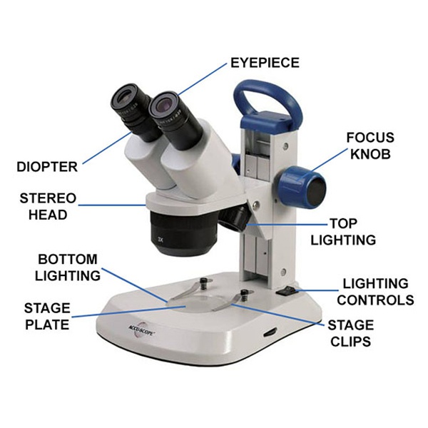
rsscience.com › stereo-microscopeParts of Stereo Microscope (Dissecting microscope) – labeled ... Labeled part diagram of a stereo microscope Major structural parts of a stereo microscope. There are three major structural parts of a stereo microscope. The viewing Head includes the upper part of the microscope, which houses the most critical optical components, including the eyepiece, objective lens, and light source of the microscope.
Platelets (Thrombocytes): Function, Normal Values, and More Platelets, also known as thrombocytes , are special blood cells that control blood clotting. Their main function is to heal a wound and stop the bleeding. A normal platelet count is 150,000-450,000 platelets per microliter. Some people have a low platelet count, which puts them at risk for uncontrolled bleeding.
Stem cell microencapsulation maintains stemness in inflammatory ... Scale bar = 50 μm. f TEM micrograph of spheroid@ [Fe III -TA]. The red dashed line indicated the area of interested magnified images. The yellow arrow indicated the cell membrane. Scale bar = 1 μm....
Dinoflagellate - Wikipedia Etymology. The term "dinoflagellate" is a combination of the Greek dinos and the Latin flagellum.Dinos means "whirling" and signifies the distinctive way in which dinoflagellates were observed to swim.Flagellum means "whip" and this refers to their flagella.. History. In 1753, the first modern dinoflagellates were described by Henry Baker as "Animalcules which cause the Sparkling Light in Sea ...
opencv windows forms c++ app bad performance on windows 11 tablet I am currently developing software, which is supposed to display and record video from 2 webcams simultaneously on a Windows 11 tablet. I've ran into a strange problem that on my main machine (i9-9900kf 16gb ram) the app works just fine, but on the actual tablet Lenovo IdeaPad Duet 3 8/128 (Intel Celeron N4020 8gb ram) it works pretty bad.
Protein Synthesis: Definition, Steps, and Diagram (2022) Protein Synthesis Diagram Translation completes in the cytoplasm of the cell and consist of four phases- (1) Activation (2) Initiation (3) Elongation (4) Termination The initiation starts with the binding of the ribosome to 5'end of mRNA. In elongation, the binding of aminoacyl-tRNA to the ribosome occurs.
BIO 240 SYLLABUS FALL 2022 2.doc - NATURAL SCIENCES... Digestive System: Anatomy 24 621 - 648 Models, Microscope slides & Dissection of Fetal pig 4. Digestive System: Physiology of Enzyme Action 24 649 - 650 Carbohydrate digestion 650 - 651 Protein digestion 652 - 653 Lipid digestion 653 - 662 5. Urinary System: Anatomy 25 663 - 688 Models, Microscope slides & Sheep kidney dissection 6.
Compound Microscope- Definition, Labeled Diagram, Principle, … 03/04/2022 · Magnification of compound microscope. In order to ascertain the total magnification when viewing an image with a compound light microscope, take the power of the objective lens which is at 4x, 10x or 40x and multiply it by the power of the eyepiece which is typically 10x.
Light Microscope (Theory) - Amrita Vishwa Vidyapeetham Microscope is an optical instrument that uses lens or combination of lens to produce magnified images that are too small to seen by unaided eye. Microscope provides the enlarged view that helps in examining and analyzing the image.














Post a Comment for "42 microscope labeled diagram"