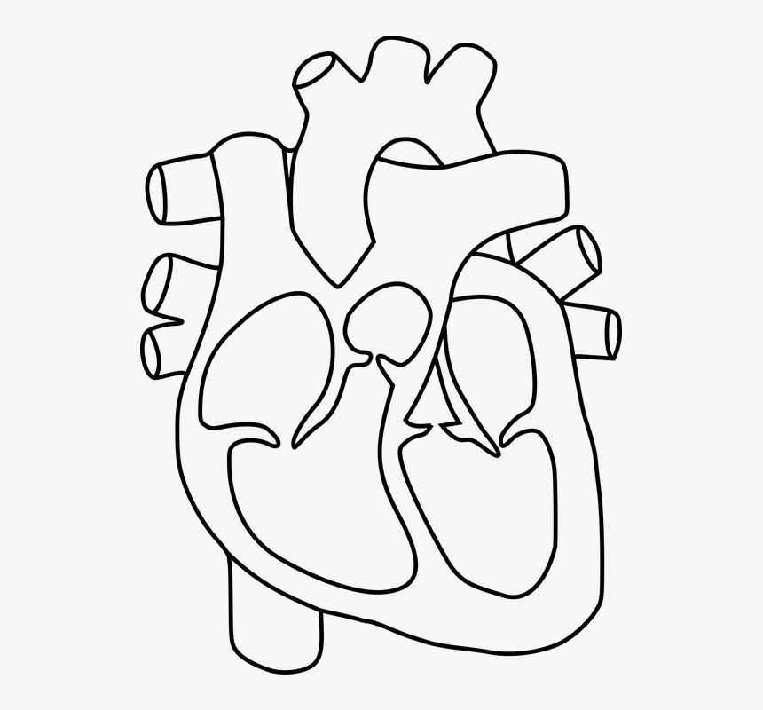
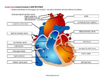
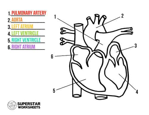
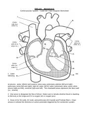
38 label the heart diagram
Labeling The Heart - GCSE Level - YouTube A revision lesson demonstrating how to label a simple diagram of the heart including the atria, ventricles pulmonary vessels and systemic vessels.This video ... Heart Anatomy: Labeled Diagram, Structures, Blood Flow ... - EZmed Anatomy of the Heart: Structures and Blood Flow [Cardiology Made Easy] Watch on Anatomy of the human heart made easy using labeled diagrams of the main cardiac structures, along with their function, blood flow through the heart, and a review with a quiz at the end to test your knowledge! Save Time with a Video!
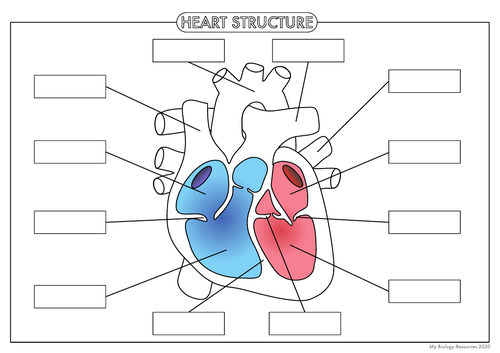
Simple Heart Diagram Labeling Activity (Teacher-Made) - Twinkl This simple heart diagram with labels activity will help your pupils begin to understand the heart, what it does and the different parts that comprise it. The resource comes with two different diagrams of the heart; one with labels attached, and one blank diagram with the labels at the bottom for students to complete themselves. Ideal as an introductory lesson on the heart and how it ...

Label the heart diagram
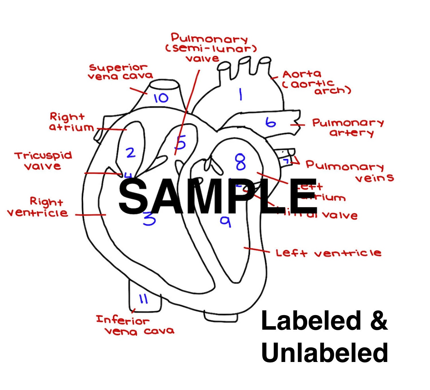
Label the Heart Diagram | Quizlet Label the Heart Diagram | Quizlet Subjects Expert solutions Log in Sign up Label the Heart 4.5 (51 reviews) + − Learn Test Match Created by bluesas9 Terms in this set (15) Superior Vena Cava ... Right Ventricle ... Left Atrium ... Atrioventricular/Tricuspid Valve ... Atrioventricular/Mitral Valve ... Septum ... Right Atrium ... Semi-lunar Valves Human Heart (Anatomy): Diagram, Function, Chambers, Location in Body The heart is a muscular organ about the size of a fist, located just behind and slightly left of the breastbone. The heart pumps blood through the network of arteries and veins called the... Label the heart - Teaching resources - Wordwall The Heart Labelled diagram by Swright Copy of Label The Diagram of The Heart Labelled diagram by Nickidocherty Copy of Heart Label Labelled diagram by Lisa148 The Heart True or false by Sfenner PE The Heart Find the match by Kmckirdyx Y10 Biology The heart Match up by Bethanyward99 Biology The Heart Labelled diagram by Scarlettfrost KS4 Biology
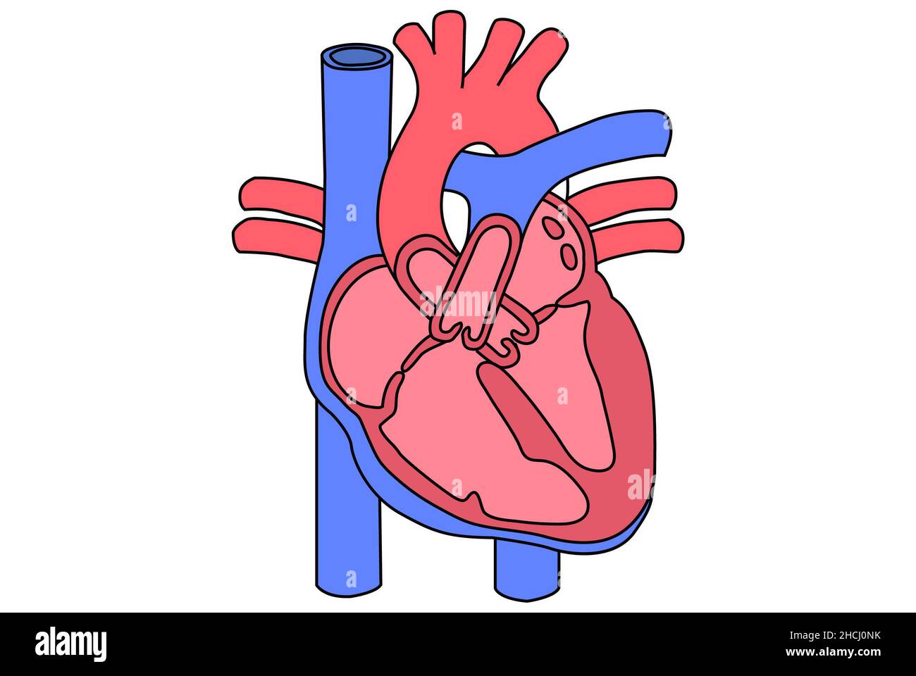
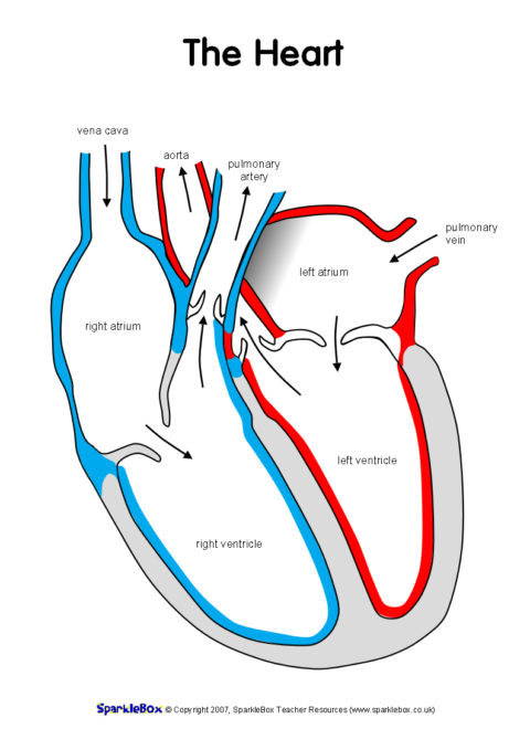
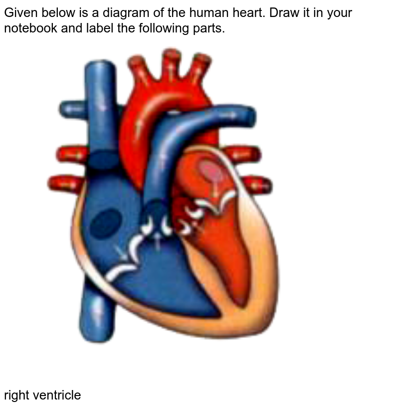
Label the heart diagram. The heart is made up of four chambers: Atria: Upper two chambers of the ... The average ...April 30th, 2018 - Template Information Title Blank Heart Diagram Labeled Categories Blank ♦ Publised Tuesday December 27th 2016 04 10 12 AM Heart Labeling Internal The Biology Corner April 29th, 2018 - Heart Worksheet The human heart is similar to the and most diagrams will show the heart as it is viewed Label each of the ... Heart Diagram for Kids - Bodytomy As you can see in the diagram of the heart, that heart is divided in four chambers, namely, right atrium, left atrium, right ventricle and left ventricle. Each of the chambers is separated by a muscle wall known as Septum. The left side of the heart receives oxygen rich blood from the lungs and pumps it out the whole body. How to Draw a Human Heart: An Easy Step-By-Step Guide - wikiHow Label the parts of the heart to reference it for anatomy. If you're trying to identify parts of the heart for a class or just for fun, consider adding the names of each segment. Neatly print the names around your drawing and then use a ruler to draw an arrow to the corresponding part. Refer to anatomical diagram to double-check your work. Label the Heart Diagram Quiz - PurposeGames.com Label the Heart Diagram by hshaw01 476 plays 18 questions ~ 50 sec More 1 too few (you: not rated) Language English Tries Unlimited [?] Last Played February 22, 2022 - 12:00 am There is a printable worksheet available for download here so you can take the quiz with pen and paper. Remaining 0 Correct 0 Wrong 0 Press play! 0% 08:00.0 Highscores
Heart Diagram - 15+ Free Printable Word, Excel, EPS, PSD Template ... Vintage Anatomical Heart Diagram. While studying anatomy heart is a major part to be covered. To study this lot of templates are available online which can be used by easily getting them online. Heart Label Interior Doc Format. services.juniata.edu | An interior format of a heart diagram has been shown in this template. This clearly shows the ... Human Heart - Diagram and Anatomy of the Heart - Innerbody The heart contains 4 chambers: the right atrium, left atrium, right ventricle, and left ventricle. The atria are smaller than the ventricles and have thinner, less muscular walls than the ventricles. The atria act as receiving chambers for blood, so they are connected to the veins that carry blood to the heart. Label the Heart Quiz - PurposeGames.com There is a printable worksheet available for download here so you can take the quiz with pen and paper. From the quiz author Ummmmmmm . . . it's pretty self explanatory . . . you label the heart. Just remember one thing - you're looking at the heart like it's in someone else so right and left are switched around. Remaining 0 Correct 0 Wrong 0 Label the Heart - Labelled diagram - Wordwall Label the Heart. Share Share by Banksm. Show More. Like. Edit Content. Embed. More. Leaderboard. Show more Show less . This leaderboard is currently private. Click Share to make it public. This leaderboard has been disabled by the resource owner. This leaderboard is disabled as your options are different to the resource owner. ...
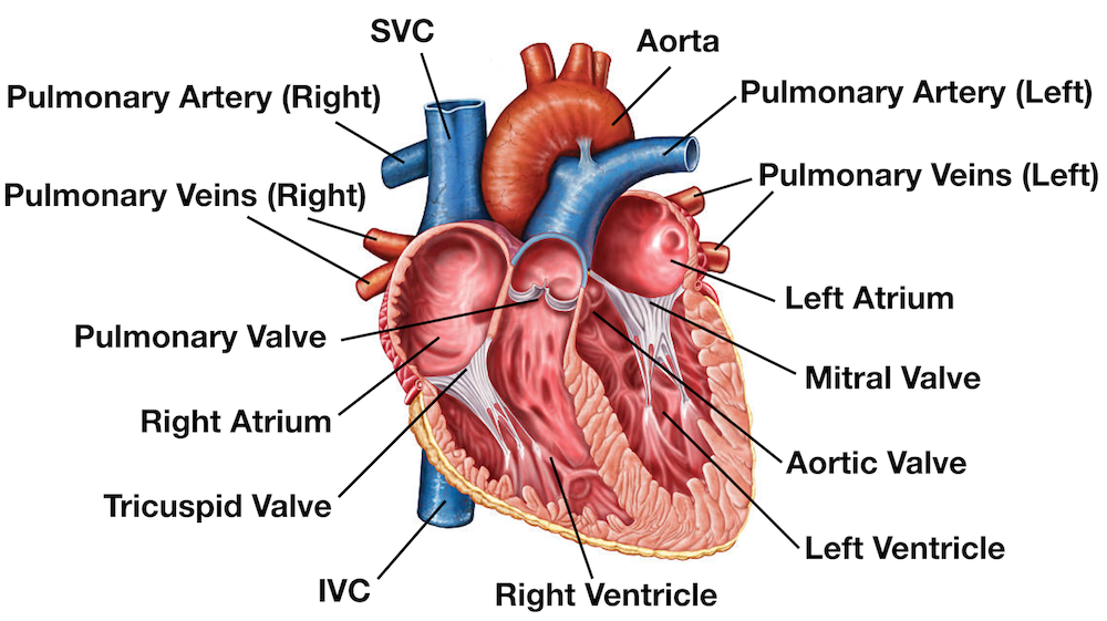
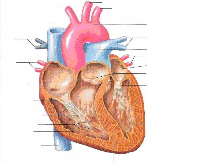
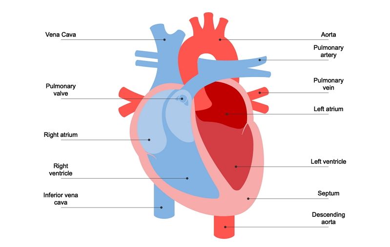
Diagram of Human Heart and Blood Circulation in It A heart diagram labeled will provide plenty of information about the structure of your heart, including the wall of your heart. The wall of the heart has three different layers, such as the Myocardium, the Epicardium, and the Endocardium. Here's more about these three layers. Epicardium Blood Flow Through The Heart: A Simple 12 Step Diagram - EZmed Step 1 and 6 involve a blood vessel, which makes sense as this is how blood enters and exits that side of the heart. Steps 2-5 involve a chamber, valve, chamber, and valve. So if you remember this general pattern, it will help you recall the order in which blood flows through each side of the heart. Human Heart Diagram Labeled - Science Trends The left ventricle and left atrium make up the left heart while the right ventricle and right atrium make up the right heart. While there are four different chambers of the heart, the chambers work together and the heart basically functions as a single organ. The muscle tissue dividing the two halves of the heart is referred to as the septum. Diagrams, quizzes and worksheets of the heart | Kenhub Labeled heart diagrams Take a look at our labeled heart diagrams (see below) to get an overview of all of the parts of the heart. Once you're feeling confident, you can test yourself using the unlabeled diagrams of the parts of the heart below. Labeled heart diagram showing the heart from anterior Unlabeled heart diagrams (free download!)
Label the heart — Science Learning Hub In this interactive, you can label parts of the human heart. Drag and drop the text labels onto the boxes next to the diagram. Selecting or hovering over a box will highlight each area in the diagram. pulmonary vein semilunar valve right ventricle right atrium vena cava left atrium pulmonary artery aorta left ventricle Download Exercise Tweet
Labelling the heart — Science Learning Hub The heart is a muscular organ that pumps blood through the blood vessels of the circulatory system. Blood transports oxygen and nutrients to the body. It is also involved in the removal of metabolic wastes. In this activity, students use online and paper resources to identify and label the main parts of the heart.
Label the HEART | Circulatory System Quiz - Quizizz The body Left ventricle Right Atrium Lungs Question 16 45 seconds Q. Where does the blood go after it goes into the pulmonary arteries? answer choices lungs heart head hands Question 17 30 seconds Q. What part of the heart delivers richly oxygenated blood to the body? answer choices Aorta Superior vena cava
The Anatomy of the Heart, Its Structures, and Functions - ThoughtCo The heart is the organ that helps supply blood and oxygen to all parts of the body. It is divided by a partition (or septum) into two halves. The halves are, in turn, divided into four chambers. The heart is situated within the chest cavity and surrounded by a fluid-filled sac called the pericardium. This amazing muscle produces electrical ...
Circulatory System Diagram - Cardiovascular System and Blood ... What is a Circulatory System Diagram. Circulatory system diagrams are visual representations of the circulatory system, also referred to as the cardiovascular system. It is comprised of three parts: the pulmonary circulation, coronary circulation, and systemic circulation. The main function of the circulatory system is to circulate blood, which ...
Heart diagram labelling quiz - ESL Games Plus Heart diagram labelling quiz When you make a closed fist, it will roughly be the size of your own heart. In addition, if you close and open your fists around 60 times per minute - the normal resting heart rate for adults - you can catch a glimpse at how hard the heart works to pump blood throughout your body. The heart is an extremely vital organ.
Unlabelled heart diagram - Healthiack Matej G. is a health blogger focusing on health, beauty, lifestyle and fitness topics. He has been with healthiack.com since 2012 and has written and reviewed well over 500 coherent articles.
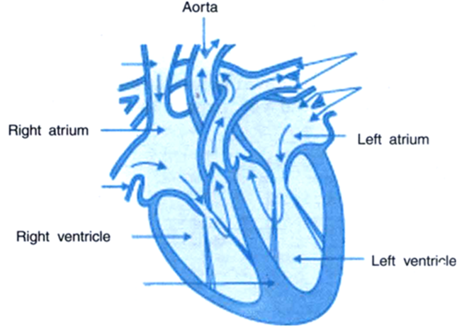
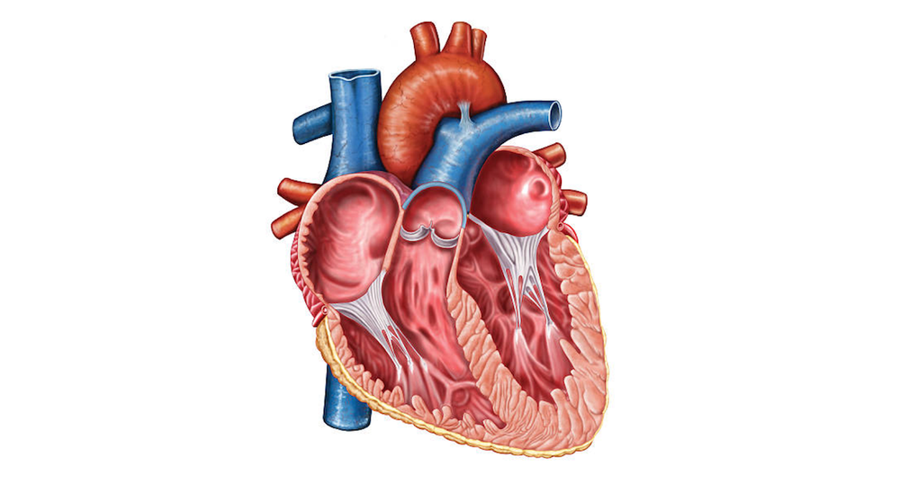
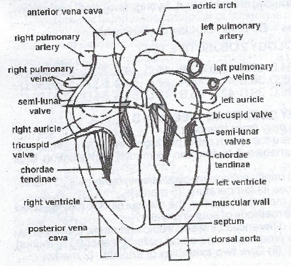
A Labeled Diagram of the Human Heart You Really Need to See The human heart, comprises four chambers: right atrium, left atrium, right ventricle and left ventricle. The two upper chambers are called the left and the right atria, and the two lower chambers are known as the left and the right ventricles. The two atria and ventricles are separated from each other by a muscle wall called 'septum'.
Heart Diagram with Labels and Detailed Explanation - BYJUS Well-Labelled Diagram of Heart The heart is made up of four chambers: The upper two chambers of the heart are called auricles. The lower two chambers of the heart are called ventricles. The heart wall is made up of three layers: The outer layer of the heart wall is called epicardium. The middle layer of the heart wall is called myocardium.
Anatomy | Label the Heart Diagram | Quizlet Anatomy | Label the Heart + − Flashcards Learn Test Match Created by justinaadoll TEACHER Terms in this set (15) Right Atrium Receives deoxygenated blood from the body Right Ventricle Pumps deoxygenated blood to the lungs Left Atrium Chamber that receives oxygenated blood from the pulmonary veins Left Ventricle Pumps oxygenated blood into the aorta
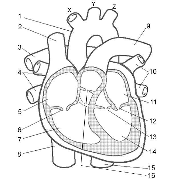
Heart Labeling Quiz: How Much You Know About Heart Labeling? Here is a Heart labeling quiz for you. The human heart is a vital organ for every human. The more healthy your heart is, the longer the chances you have of surviving, so you better take care of it. Take the following quiz to know how much you know about your heart. Questions and Answers 1. What is #1? 2. What is #2? 3. What is #3? 4. What is #4?
Label the heart - Teaching resources - Wordwall The Heart Labelled diagram by Swright Copy of Label The Diagram of The Heart Labelled diagram by Nickidocherty Copy of Heart Label Labelled diagram by Lisa148 The Heart True or false by Sfenner PE The Heart Find the match by Kmckirdyx Y10 Biology The heart Match up by Bethanyward99 Biology The Heart Labelled diagram by Scarlettfrost KS4 Biology
Human Heart (Anatomy): Diagram, Function, Chambers, Location in Body The heart is a muscular organ about the size of a fist, located just behind and slightly left of the breastbone. The heart pumps blood through the network of arteries and veins called the...
Label the Heart Diagram | Quizlet Label the Heart Diagram | Quizlet Subjects Expert solutions Log in Sign up Label the Heart 4.5 (51 reviews) + − Learn Test Match Created by bluesas9 Terms in this set (15) Superior Vena Cava ... Right Ventricle ... Left Atrium ... Atrioventricular/Tricuspid Valve ... Atrioventricular/Mitral Valve ... Septum ... Right Atrium ... Semi-lunar Valves















Post a Comment for "38 label the heart diagram"