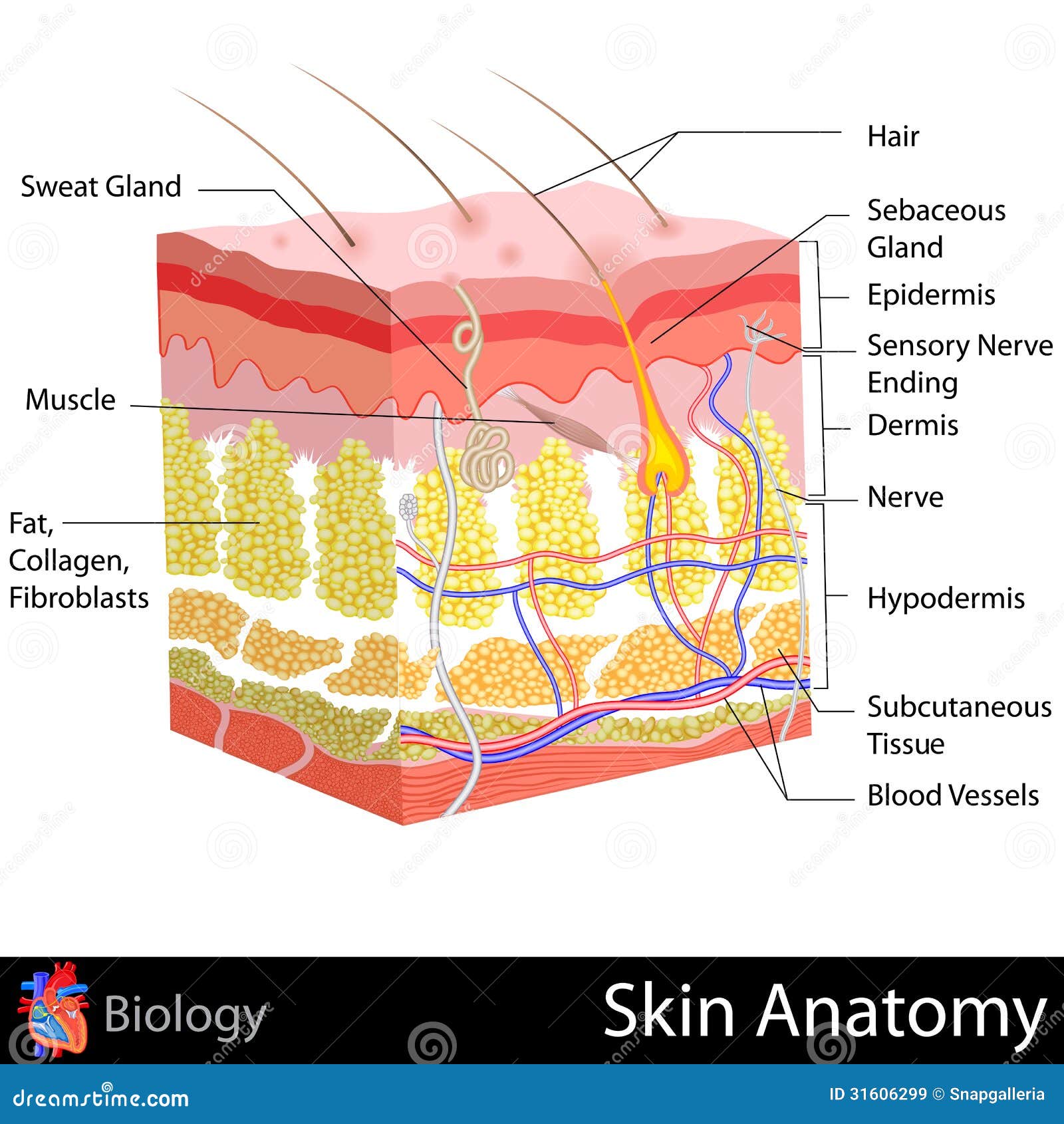
39 skin diagram with labels
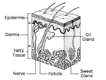
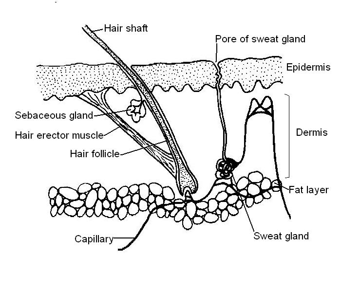
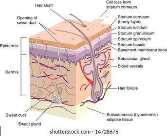
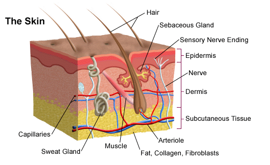
Simple diagram of the skin - Healthiack Simple diagram of the skin This brief article is displaying Simple diagram of the skin … Please click on the diagram (s) to view larger version. You're welcome to browse healthiack.com for more details on this specific topic. Best viewed on 1280 x 768 px resolution in any modern browser. Simple diagram of the skin 1285 Labeled diagram of the skin & skin stem cells in research Labeled diagram of the skin epidermis. Source, Professor Paul Knoepfler, UC Davis School of Medicine. Labeled diagram of the skin: epidermis, skin stem cells I've included another labeled diagram of skin here with specific annotations for important epidermal layers. This is more zoomed in than the first figure at the top of the post.
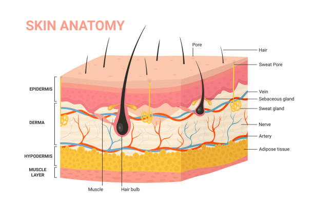
Skin Diagram Pictures, Images and Stock Photos - iStock Layer of Healthy Human Skin in vector style and components information. Illustration about medical diagram.

Skin diagram with labels
skin labeling Diagram | Quizlet Dermis Middle layer of the skin; contains collagen; location of sweat & sebaceous glands and nerve endings and capilaries Epidermis outermost layer of the skin;composed of squamous epithelium; contains keratin subcutaneous layer Innermost layer of skin, contains fat and is the location of main blood vessels basal layer of epidermis Layers of Skin: How Many, Diagram, Model, Anatomy, In Order - Healthline skin fragility syndrome boils nevus (birthmark, mole, or "port wine stain") acne melanoma (skin cancer) keratosis (harmless skin growths) epidermoid cysts pressure ulcers (bedsores) Dermis... 5.1 Layers of the Skin - Anatomy & Physiology "Thick skin" is found only on the palms of the hands and the soles of the feet. It has a fifth layer, called the stratum lucidum, located between the stratum corneum and the stratum granulosum (Figure 5.1.2). Figure 5.1.2 - Thin Skin versus Thick Skin: These slides show cross-sections of the epidermis and dermis of (a) thin and (b) thick ...
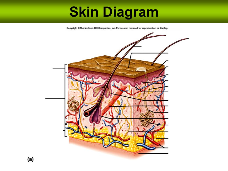
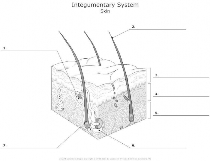
Skin diagram with labels. PDF Skin Diagram Labeling - New Providence School District Skin Diagram Labeling . 1. Label the diagram with the . letters. below according to the structure/area they describe. You may label with a line or put the label directly onto the area described. Be as precise as possible. If you are worried about the precision of your label add a word after to explain exactly where your label should be. Skin Diagram | Worksheet | Education.com Skin Diagram. Though skin may seem like nothing more than the source of things like zits and oil to your pre-teen, skin actually has many important jobs to do. Learn more about the skin (and the science behind pimples -- ew!) in this printable life science diagram. Skin diagram - Teaching resources - Wordwall Skin diagram Examples from our community 10000+ results for 'skin diagram' Skin Diagram Labelled diagram by U51678338 Skin Diagram Labelled diagram by Laurabrown1 Skin diagram Labelled diagram by Siobhan45 Skin diagram Labelled diagram by Sambrackley Skin diagram Labelled diagram by Jordannao Skin diagram to label Labelled diagram by Kmiller14 The Skin (Human Anatomy): Picture, Definition, Function, and Skin ... The skin is the largest organ of the body, with a total area of about 20 square feet. The skin protects us from microbes and the elements, helps regulate body temperature, and permits the...
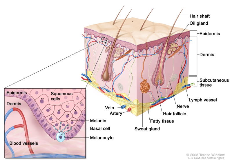
Skin diagram labeled - Healthiack Skin diagram labeled By Matej Gololicic 10 SHARES Skin diagram labeled This brief article displays Skin diagram labeled … Please click on the diagram (s) to view larger version. You are welcome to browse healthiack.com for more details on this very topic. Best viewed on 1280 x 768 px resolution in any modern browser. Skin diagram labeled 1075 Labeled Skin Diagram Pictures, Images and Stock Photos Labeled Skin Diagram Stock Photos, Pictures & Royalty-Free Images - iStock Pricing Boards Sign in Join Video Back Videos home Curated sets Signature collection Essentials collection Diversity and inclusion sets Trending searches Video Alabama tennessee Mexico bar Mexico Jalin hyatt Armadillo Selfie Astronaut Realm to Tennessee Academy museum gala Skin Anatomy Cross Section with Labels on White stock photo - iStock Jan 12, 2018 ... Labeled medical diagram, a 3D cross section of human skin layers and parts such as a hair follicle and sweat glands on a white background. Anatomy of the Skin - Stanford Children's Health The skin is made up of 3 layers. Each layer has certain functions: Epidermis. Dermis. Subcutaneous fat layer (hypodermis) ...
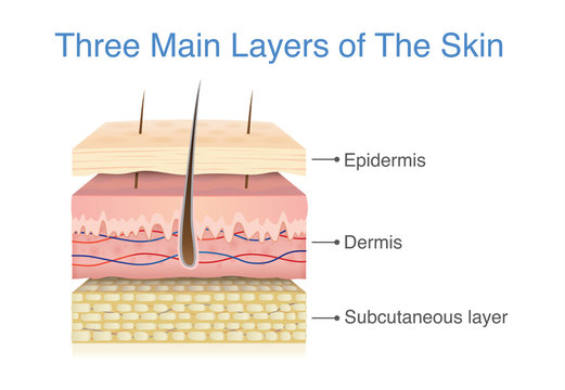
Skin 1: the structure and functions of the skin - Nursing Times Nov 25, 2019 ... Abstract Skin diseases affect 20-33% of the population at any one time, and around 54% of the UK population will experience a skin condition ... Layers of the Skin | Anatomy and Physiology I - Lumen Learning The skin is composed of two main layers: the epidermis, made of closely packed epithelial cells, and the dermis, made of dense, irregular connective tissue that houses blood vessels, hair follicles, sweat glands, and other structures. Beneath the dermis lies the hypodermis, which is composed mainly of loose connective and fatty tissues. Skin Labeling | Biology Game | Turtle Diary Skin Labeling - Biology Game Identify and label figures in Turtle Diary's interactive online game, Skin Labeling! Drag the given words to the correct blanks to complete the labeling! label the skin diagram — Printable Worksheet - PurposeGames.com This is a free printable worksheet in PDF format and holds a printable version of the quiz label the skin diagram. By printing out this quiz and taking it with pen and paper creates for a good variation to only playing it online. Other Quizzes Available as Worksheets label the digestive system Medicine Creator k8e1404 Quiz Type Image Quiz Value
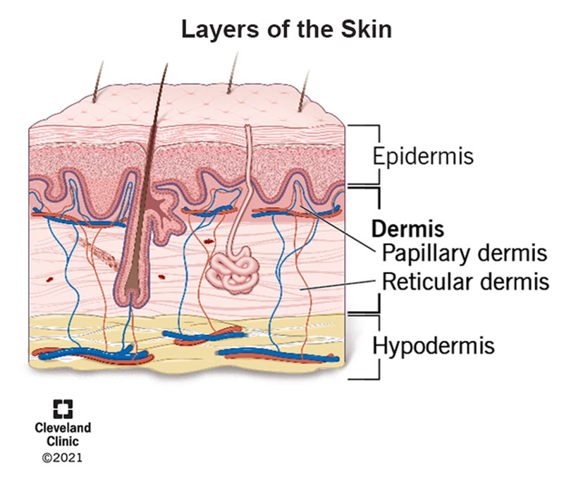
Skin Layers: Structure, Function, Anatomy, and More - Verywell Health There are three main layers of skin: Epidermis: The outermost layer, which contains five sub-layers Dermis: The middle layer, which consists of two parts known as the papillary dermis (thin, upper layer) and the reticular dermis (thick, lower layer) Subcutaneous tissue: The deepest layer of skin What is the integumentary system?
How to draw skin LS - Pinterest Apr 13, 2016 - Step by step tutorials on drawing biology diagrams. ... The skin consists of two main layers called epidermis and dermis.
Skin Diagram Labeling Skin Diagram Labeling. 1. Label the diagram with the letters below according to the structure/area they describe. You may label with a line or put the label ...
Anatomy, Diagram and Function of Skin - VEDANTU The skin has a surface area of between 16.1-21.5 sq ft. for an adult human. The thickness of the skin differs over all parts of the body, and between men and women and the young and the old. For example, the skin on the forearm which is on average 1.3 mm in the human male and 1.26 mm in the human female.
Skin Diagram Labeled Pictures, Images and Stock Photos Skin Diagram Labeled Pictures, Images and Stock Photos View skin diagram labeled videos Browse 38 skin diagram labeled stock photos and images available, or start a new search to explore more stock photos and images. Newest results Systemic lupus erythematosu Systemic lupus erythematosu. SLE or lupus, is a systemic autoimmune disease.
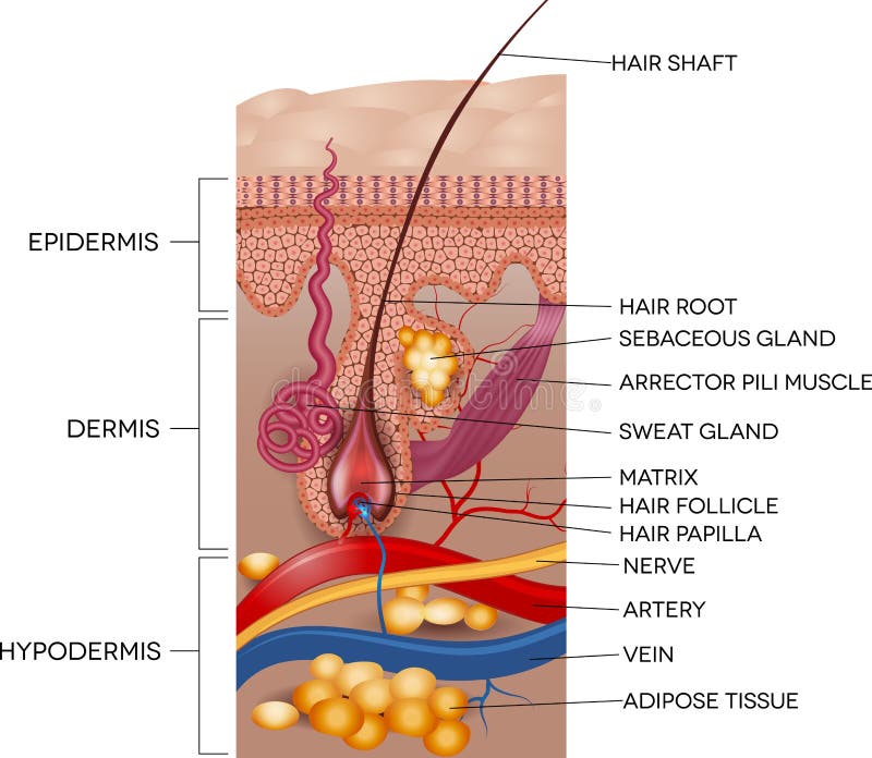
Labeled Skin and Hair Anatomy Stock Vector - Dreamstime Illustration about Labeled Skin and hair anatomy. Detailed medical illustration. Illustration of diagram, labeled, layers - 49872752.
Labeled Skin Structure Diagram | Quizlet Dermis Fibrous and elastic tissue, provides strength and elasticity to the skin and supports the epidermis, home to hair follicles, glands, nerves etc Papillary Layer Upper dermal layer, provides the epidermis with nutrients and regulates body temperature Reticular Layer
Diagram of human skin structure — Science Learning Hub Diagram of human skin structure. Image. Add to collection. Tweet. Rights: University of Waikato Published 1 February 2011 Size: 100 KB Referencing Hub media. The epidermis is a tough coating formed from overlapping layers of dead skin cells.
Skin Diagram Teaching Resources | Teachers Pay Teachers Integumentary System: Skin Diagram to Label by Lori Maldonado 4.9 (30) $2.50 PDF Students will read the definitions and label the skin anatomy diagram. Answer key included. This diagram had been modified from Enchanted Learning. I have used this worksheet as an in-class assignment as well as a homework assignment.
Skin structure diagram - Teaching resources - Wordwall Skin Diagram 2022 Labelled diagram by Jordannao Skin Diagram 2 Labelled diagram by Jordannao Structure of the skin Labelled diagram by Lfoster1 Structure of the skin Labelled diagram by Claredorotiak Hair and Skin Structure Labelled diagram by Janehughes Skin structure - label Labelled diagram by Kellyrandall Structure of the skin Labelled diagram
label the skin diagram Quiz - purposegames.com label the skin diagram by k8e1404 89 plays 12 questions ~ 30 sec More 0 too few (you: not rated) Language English Tries 12 [?] Last Played February 22, 2022 - 12:00 am There is a printable worksheet available for download here so you can take the quiz with pen and paper. Remaining 0 Correct 0 Wrong 0 Press play! 0% 08:00.0 Highscores

Human Skin Anatomy Cross Section Diagram Chart Art Print Stand or Hang Wood Frame Display Poster Print 13x9
Integumentary system parts: Quizzes and diagrams | Kenhub Spend some time analyzing the skin diagram labeled above. Try to memorize the appearance and location of each structure. Learning the function of each structure will accelerate your ability to memorize, so be sure to check out our detailed article on The Integumentary System parts and functions. ...
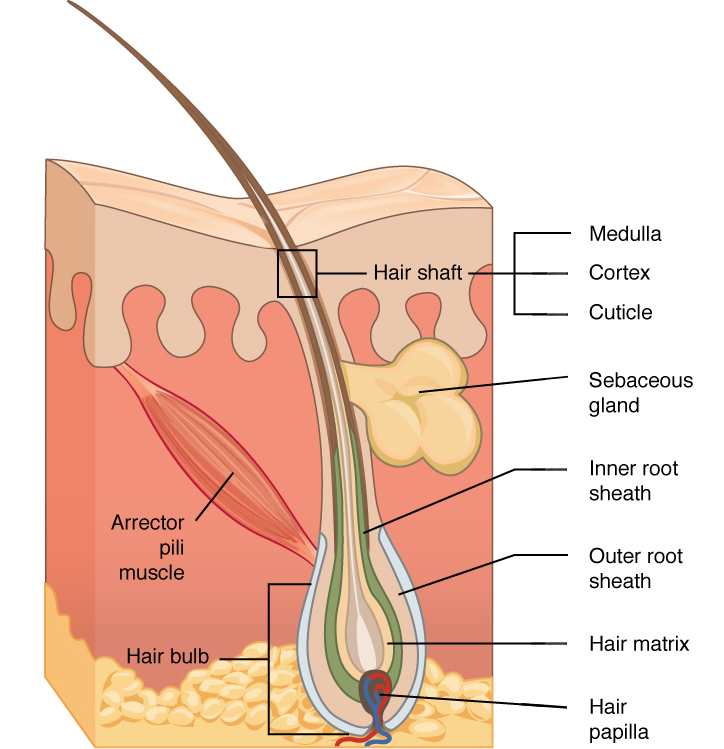
Skin Histology Slide Identification - AnatomyLearner I hope these skin microscope slide labeled diagrams might help you to identify and learn all the structures. If you need more skin microscope slide labeled diagram, please follow anatomy learner on social media. I will update or upload a new skin slide labeled diagram on social media (if any correction). Functions of skin
Skin Diagram || How to draw and label the parts of skin The skin consists of two main layers called epidermis and dermis. Epidermis is the layer of protection. It has sweat pores and small hairs. Dermis lies below the epidermis. It is made up of...
A Human Body Skin-structure Quiz! - ProProfs Quiz Start. Create your own Quiz. In this, a human body skin structure quiz, we are going to focus on the underlying and the most elementary structure of the human body. It's easy to take your skin for granted, but when you consider how it protects your body from harm, it is something we should appreciate more.
Skin Diagram || How to draw and label the parts of skin - Pinterest Mar 28, 2021 - 'Skin Diagram || How to draw and label the parts of skin' is demonstrated in this video tutorial step by step.The sense of touch had received ...
A diagrammatic representation of the structure of human skin in cross... Diagram is not to scale. from publication: Skin Deep: The Basics of Human Skin ... used in dermatology for treating inflammatory diseases, was spin labeled ...
Human Skin Diagram & Function | How Many Layers of Skin Are There ... There are seven layers to the human skin. In order from superficial to deep (external to internal) they are 1. Stratum corneum 2. Stratum lucidum 3. Stratum granulosum 4. Stratum spinosum 5....
Skin Diagram with Detailed Illustrations and Clear Labels - BYJUS Skin Diagram with Detailed Illustrations and Clear Labels Biology Important Diagrams Skin Diagram Skin Diagram The largest organ in the human body is the skin, covering a total area of about 1.8 square meters. The skin is tasked with protecting our body from external elements as well as microbes. Interesting Note:
Label Skin Diagram Printout - EnchantedLearning.com Read the definitions, then label the skin anatomy diagram below. blood vessels - Tubes that carry blood as it circulates. Arteries bring oxygenated blood from the heart and lungs; veins return oxygen-depleted blood back to the heart and lungs. dermis - (also called the cutis) the layer of the skin just beneath the epidermis.
5.1 Layers of the Skin - Anatomy & Physiology "Thick skin" is found only on the palms of the hands and the soles of the feet. It has a fifth layer, called the stratum lucidum, located between the stratum corneum and the stratum granulosum (Figure 5.1.2). Figure 5.1.2 - Thin Skin versus Thick Skin: These slides show cross-sections of the epidermis and dermis of (a) thin and (b) thick ...
Layers of Skin: How Many, Diagram, Model, Anatomy, In Order - Healthline skin fragility syndrome boils nevus (birthmark, mole, or "port wine stain") acne melanoma (skin cancer) keratosis (harmless skin growths) epidermoid cysts pressure ulcers (bedsores) Dermis...
skin labeling Diagram | Quizlet Dermis Middle layer of the skin; contains collagen; location of sweat & sebaceous glands and nerve endings and capilaries Epidermis outermost layer of the skin;composed of squamous epithelium; contains keratin subcutaneous layer Innermost layer of skin, contains fat and is the location of main blood vessels basal layer of epidermis















Post a Comment for "39 skin diagram with labels"